
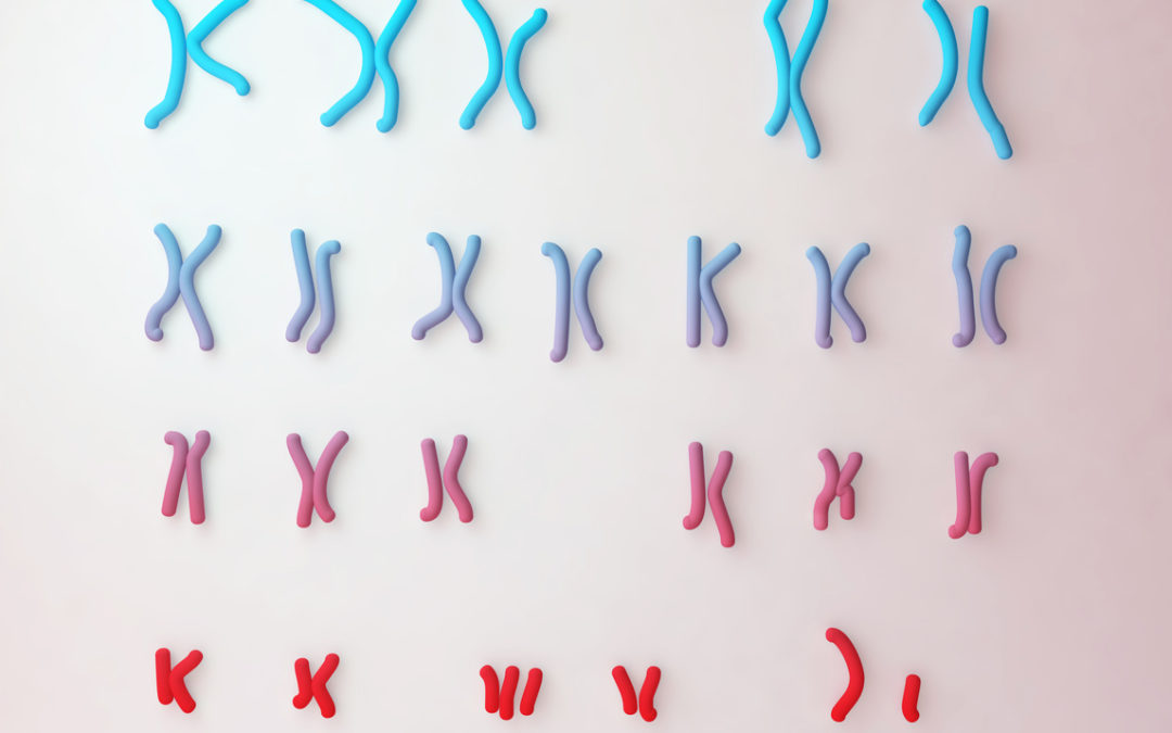
For patients undergoing in vitro fertilization (IVF), preimplantation genetic testing for aneuploidy (PGT-A) can be offered to screen their developing embryos for chromosomal abnormalities prior to implantation. This technology may decrease the time to achieve a successful pregnancy for some patients. PGT-A is a rapidly changing technology, which has improved since its inception in 1990. Researchers continue to refine the technique to enhance the safety and accuracy of the testing.
Traditionally, PGT is performed by collecting a small number of cells for testing from the embryo about 5 days after fertilization. At this time, the embryo has two groups of cells. The “trophectoderm” is a ring of cells around the outside edge of the embryo. These cells do not form the fetus, but go on to develop into the placenta of the pregnancy. The part of the embryo that will become the fetus is a cluster of cells inside of this ring, called the “inner cell mass.” Traditionally, cells biopsied as part of the PGT process are taken from the trophectoderm because it generally shares the same DNA, or chromosomal makeup, as the inner cell mass.
Some scientists and doctors have questioned whether PGT by trophectoderm biopsy could harm the developing embryo, leading to fewer viable embryos or potential maternal and fetal risks for ongoing pregnancies. While most of the available data is reassuring, more information is needed to clarify whether this biopsy process could truly increase risks. Another concern is that the trophectoderm is sometimes not genetically identical to the inner cell mass. This “mosaicism” can occasionally create false positive and false negative PGT results.
Due to concerns about the traditional PGT trophectoderm biopsy process, new research is investigating a “non-invasive” method of gathering DNA needed for PGT. Non-invasive preimplantation genetic testing (niPGT) utilizes the same DNA testing technology as traditional PGT, but it does not require removal of cells from the embryo. Scientists have learned that as embryos grow in the laboratory, they release small amounts of their DNA into the surrounding fluid, or “culture media,” in which they are grown. The idea behind niPGT is to test this DNA in the media instead of removing cells from the embryo itself.
The technique was first published in 2016 when researchers identified enough DNA in the culture media of several embryos to provide PGT results; however, false positives and false negatives were observed in this small study. More recent data suggests that niPGT is a promising technology, theoretically able to detect the chromosomal makeup of both the trophectoderm and the inner cell mass. If niPGT is able to provide a more accurate representation of the chromosomal make-up of the inner cell mass, then it may be as accurate or more accurate than current PGT methods without the potential risks of the biopsy. However, much is unknown about this new technology. The source of the DNA in the media is not completely understood at this time, so it cannot be proven that the DNA is derived from both the trophectoderm and the inner cell mass. Large studies will be needed to validate this new technology further before a complete understanding of its accuracy can be gained.
Given the experimental nature of niPGT, this new technology is not yet widely available in a clinical setting. The hope is that future studies will help clarify whether niPGT may be a more successful alternative to traditional PGT. If you are considering IVF with PGT, be sure to discuss the available embryo testing options with your doctor or genetic counselor to determine what reproductive option may be best for you.

Elysia is an independent consultant to Sharing Healthy Genes. She is a board-certified Reproductive Genetic Counselor and Clinical Instructor at the University of North Carolina in Chapel Hill. She received her Bachelor’s in Science at the University of Maryland in College Park and her Master’s in Genetic Counseling from the University of Maryland in Baltimore. In her role as educator, Elysia is involved in didactic and clinical teaching of a variety of learners, including medical students, residents, and fellows, as well as genetic counseling students. As a clinician, Elysia provides genetic counseling services for patients considering pregnancy and who are currently pregnant. Elysia has enjoyed a number of leadership roles in professional committees with the National Society of Genetic Counselors, the American Board of Genetic Counseling, and the Accreditation Council for Genetic Counseling.